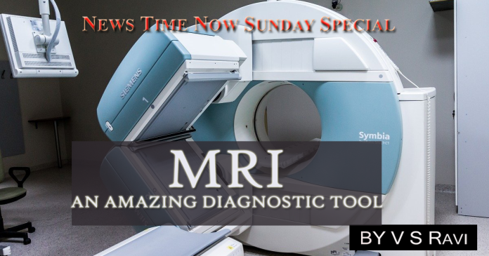
The last few decades have witnessed a revolution in medical diagnosis with the help of extremely sophisticated techniques. In the 1970s the CAT (Computerised Axial Tomography) scan had become operational enabling physicians to make cross section X- rays of the human body. CT images which can be compared to slices of a bread produced by a knife can discern only the density of anatomical structures. Another scanner called PETT, introduced a little later, enables detection of chemical reactions in the body. The PETT scanner does not use X-rays but measures emissions from a radioactive isotope injected into the blood.
However, both CT and PETT use ionising radiation, high doses of which cause cancer and infertility. Fortunately though both methods require only very low doses of radiation, the exact level of radiation that would push a patient to the danger zone has not been estimated. On the other hand, a radically new tool known as the Nuclear Magnetic Resonance Scanner (which produces Magnetic Resonance Images) does not use X-rays or any other material that exposes patients or operators to dangerous ionising radiation but merely uses harmless magnets and a radio frequency energy millions of times weaker than that of visible light. It has revolutionised medical diagnostic procedure, like no other diagnostic procedure, including CAT scan or PET Scan.
NMR senses the exchange of atoms between molecules in physical reactions and can thus literally observe physiological processes such as the flow of blood, the shrinking of an arthritic knee after steroid therapy, the molecular changes in a muscle as it becomes fatigued due to exercise, and the behaviour of the brain as it throbs in rhythm with the heart beat or the changes in the brain after a Seizure. Its main advantage is that all this can be done without causing the patient any harm or discomfort.
NMR machines are now available in the market in almost all countries. Many reputed hospitals and clinics all over the world are already using this ‘miracle’ machine for medical investigation.
The idea of NMR basically sprang out of laboratory method that had been used by chemists since the 1940s to analyse small chemical samples by exposing them to magnetic fields. A Brooklyn doctor had predicted in 1973 that this technique can be used for taking pictures of nuclei inside the body. In 1978 EMI Limited, the British company that had pioneered the CT machine produced an NMR scan of human head. In 1981, a team of scientists in Scotland published NMR images in two medical journals showing that the method could distinguish normal tissue from abscesses and tumours inside patients. Since other interesting results have been reported concerning the use of NMR.
At a hospital in London about 300 patients and 50 healthy volunteers had been given both NMR scans and CT scans. In 10 patients with multiple sclerosis (a disease in which the fatty sheaths surrounding nerve fibres wither away and are replaced with hard plaques), CT scans revealed only 19 plaques whereas NMR process revealed 110. Some doctors are confident that NMR can be used to diagnose diseases such as multiple sclerosis and monitor their course.
By about the middle of 1982 about a thousand patients and volunteers in the United States and England had been subjected to NMR scans, with no consequent after-effects. In Scotland one radiologist scanned patients with rheumatoid arthritis and was able to detect the swelling in their knee that began after the doctor had injected a ‘contrast medium’ a substance that makes X-ray images clear.
Following steroid treatment, NMR scan revealed the shrinking of these tissues. NMR scans not only indicated that the contrast medium caused swelling but also provided evidence that NMR can accomplish the same job as X-rays without the use of substances that cause irritation to tissues. Doctors are hopeful that NMR scans would prove useful in detecting a variety of other problems such as swelling of the brain, disc complications in spine, aneurysms in blood vessels and even cancer.
NMR works by activating molecules rather than breaking them apart. It measures the nuclei of elements that have an odd number of protons and neutrons and thus spin about their own axes. Nuclei have an external charge which creates a small magnetic field as they spin. NMR spectroscopy involves subjecting samples of chemicals to an external magnetic field which causes the nuclei to line up in the direction of the field. When the nuclei spin on their own axes, the axes begin to gyrate around the axes of the magnetic field in a kind of motion known as ‘precession’.
This ‘precession’ is disturbed by NMR which applies a second magnetic field at right angles to the first. That causes some of the nuclei to flip and precess at an angle perpendicular to the first field. When the second field is turned off the nuclei return to their previous positions, giving off electromagnetic signals that can be detected.
The NMR computer calculates the density of the nuclei from the time taken by the nuclei to assume their earlier alignment which is affected by the nuclei nearby. This information can then be transformed into a cross-section image. Unlike the CT scan which reveals only location and size of a structure, NMR can identify the contents also.
Today’s NMR scanners concentrate on the hydrogen atom which is abundant in the body accounting for 70 per cent of the body weight mostly in the form of water and fat. However, in order to study the biochemistry of metabolic events in the body, doctors welcomed the arrival of the next generation of scanners which can detect phosphorus, a chemical that the body passes back and forth during reactions between two different kinds of molecules.
Phosphorus occurs naturally in two isotopes of which P31 is much rarer and accounts for only one in 100 atoms of phosphorus. (Isotopes P30 and P31 represent different atomic forms of the same elements.) NMR can detect only the rarer isotope whose nucleus contains an odd number of protons and neutrons. But detection of the comparatively rare isotope P31 in a large mass such as the human body is very tricky and more advanced engineering techniques are required before it can become practical.
P31 scanners are already in use in many parts of the United States. Using such a scanner, a biochemist studied the chemical changes in a muscle during and after exercise. NMR can reveal the chemical changes that define the point beyond which a fatigued muscle cannot go without pain. For example, one biochemist had found abnormalities in a worker. However, NMR studies revealed that he in fact lacked an important enzyme and hence he reached a stage of muscle fatigue quite easily. Similarly, NMR has revealed other interesting facts to scientists in England. Previous speculation that the brain must throb with surges of blood from the heart has been proved correct by the findings of a superfast NMR scanner that^ takes a slice in 0.03 second. Physicians hope to use such an ability of the NMR scanner to diagnose diseases in the brain in future.
Scientists feel that it is desirable for doctors to have an adequate knowledge of physiology, physics and cell biology and also much greater expertise than is needed for X-ray and CT scans for using NMR. It is necessary that these opinions are taken seriously by the medical community as NMR has become available all over the world now.
MRI scanning is regularly being done, in almost all important hospitals throughout India. MRI represents one of the greatest advances in medical diagnostic procedure.


